"*" indicates required fields


Craniosynostosis in Dallas, TX
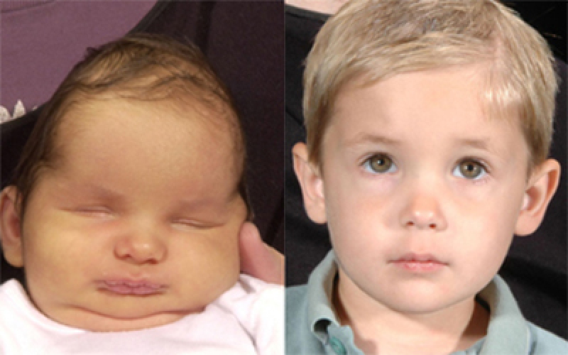
Craniosynostosis is a condition that can greatly affect a child’s future development. Recognized as world leaders in craniofacial conditions since 1971, the International Craniofacial Institute is on the cutting edge of advances in the medical industry that provide high rates of success for children affected by this condition.
What Is Craniosynostosis?
A baby’s skull is comprised of separate bones connected by sutures, rather than fused bone. These sutures normally remain open to allow the skull to expand as the brain grows. Once a child’s brain reaches its full physical size, the bones gradually fuse together, forming a solid skull. If any (or all) of the sutures close prematurely, the skull and brain cannot grow/form as they usually would, resulting in what is known as craniosynostosis.
Craniosynostosis can occur along a single type of suture line or several suture lines. The amount of variance from an average, “normal” skull shape depends on which suture(s) fused early – restricting brain growth in its programmed direction. The brain grows in the direction of least resistance. If its usual “path” is restricted, it will push the skull into an abnormal shape.
What Are the Categories of Craniosynostosis?
There are five major “categories” of craniosynostosis, designated by the suture(s) involved.
- Metopic Synostosis
- Coronal Synostosis
- Sagittal Synostosis
- Lambdoidal Synostosis
- Multiple Suture Synostosis
Craniosynostosis may be expressed as a skull-shape condition (with no other physical anomalies), as part of a craniofacial syndrome, or as a single aspect of a variety of distinct syndromes.
Syndromal craniosynostosis is a closure of multiple types of skull sutures, producing a constellation of symptoms and signs that collectively characterize the appearance and abnormal growth of the head and face. Some specific examples of syndromal craniosynostosis are Apert, Crouzon, Pfeiffer, Carpenter, and Saethre-Chotzen syndromes.
The Causes of Craniosynostosis
During early development in the womb, a baby’s FGFR-2 protein is supposed to direct immature cells to become bone cells. This protein also tells immature cells when to stop becoming bone cells. However, in craniosynostosis, we believe the FGFR-2 gene may not produce the FGFR-2 protein properly, so that it doesn’t know when to stop telling tissue to become bone. Without clear, “stop bone production” information, the soft sutures of the skull form fused bone before they should – leading to abnormal growth patterns.
This bone-formation disturbance may be the only evident concern or may lead to other difficulties. Craniosynostosis may be expressed as a skull-shape condition with no other physical anomalies, as part of a craniofacial syndrome (such as Apert, Crouzon, Muenke, or Pfeiffer), or as a single aspect of a variety of distinct syndromes.
Craniosynostosis Treatment
When a child is born with a craniofacial condition, we evaluate many factors to develop the most effective treatment plan. We study how the condition has affected the child’s underlying structures and functions, including the brain and facial skeleton, the central nervous system, the senses, and parts of the spine (cervical vertebrae). In patients whose facial skeleton is affected, we carefully identify resulting changes in the soft tissues of the face, mouth, and pharynx (top of the throat area). It’s imperative to quickly determine how the condition is affecting normal, critical functions such as breathing, swallowing and speaking so we can promptly begin appropriate treatment.
Our treatment goals are to give the child’s brain room to grow properly – in all directions without resistance – by separating or “releasing” the fused skull sutures.
Each child’s case is unique and is treated as such. Generally speaking, there are two surgical approaches to releasing sutures. We may apply a traditional, open-skull technique (cranial vault resection), or the more recently developed, less invasive endoscopic-guided surgery.
Traditional surgery involves operating for three to seven hours to perform the following:
- carefully planned ear-to-ear scalp incision
- gentle pull-back of the scalp to allow access to the fused area
- series of tiny snips to open the closed suture
- reshaping of the head to correct any skull and/or facial anomalies, replacing bone with appropriate material
- closure of the incision
- blood transfusion to replace blood loss
- recovery in the hospital for three to five days
Minimally invasive, endoscopic-guided surgery involves operating for approximately one hour to perform the following:
- one or two one-inch incisions
- insertion of the endoscope into the incisions
- location of the fused area(s)
- release of the fused area(s)
- closure of the incision(s)
- blood transfusion to replace blood loss (not required in most cases)
- recovery in the hospital for, typically, one day
- custom-fitting a “molding” helmet to direct balanced skull growth
This surgery can be conducted on babies as young as three months old up until about six months of age.
A surgeon will advise as to which type of surgery is best suited for a particular child, considering his or her type of craniosynostosis, overall health, age, etc.
Why Choose International Craniofacial Institute?
Since its founding by Dr. Kenneth Salyer in 1971, the International Craniofacial Institute has gained worldwide recognition as being one of the most advanced facilities in the treatment of craniofacial disorders. The complexity of the field means we understand our patients have an array of dental, medical, psychosocial, and surgical needs. To address these issues, our team consists of many dedicated professionals who represent more than 15 disciplines of specialty. Our physicians have successfully treated more than 17,000 patients from more than 30 different countries. We additionally train surgeons from many world locations in the advanced techniques required for craniofacial diagnosis, surgical correction, and repair.
If you have a child or another family member who is suffering from a genetic syndrome or has a cleft lip, cleft palate, or craniofacial complication, the staff at the International Craniofacial Institute can help. Contact us today to talk with the doctors and staff about your options and how we can help.


