"*" indicates required fields


Treacher Collins in Dallas, TX
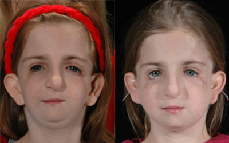
Treacher Collins syndrome or mandibulofacial dysostosis involves abnormal growth within different areas of the face. The specialists at International Craniofacial Institute in Dallas, Texas can accurately diagnose the condition and take appropriate steps toward a successful and well-coordinated treatment.
What Is Treacher Collins Syndrome?
Treacher Collins syndrome is described as a congenital disorder that presents itself with distinct craniofacial deformities. The process begins during the embryonic stage of the child’s life and often can be detected before the infant is born. The disorder is rare, and, in many cases, inherited, but in some, it is developed spontaneously without either one of the parents being affected.
How Do People Inherit Treacher Collins Syndrome?
About 60 percent of the cases result from new gene mutations. The remaining 40 percent inherits the disease when one or both parents carry the mutated gene, but do not show any signs or symptoms of the condition. The pattern of inheritance shows that each of the following generations affected with Treacher Collins syndrome face increased clinical signs and symptoms.
How Common Is Treacher Collins Syndrome?
The disorder is very unique and is estimated to affect one in 50,000 live births.
Treacher Collins Syndrome Characteristics
- Flat, underdeveloped or missing cheekbones and chin
- Disfigured or missing ears, missing ear canals
- Cleft or high vaulted palate
- Facial clefts
- Overgrowth of hair into the facial area
- Slanted downward eyes
- Orbital area abnormalities
- Missing or sparse eyelashes
- Displaced or missing TMJ
- Facial muscle deficiency
- Anterior open bite or other malocclusion
What Is the Cause of Treacher Collins Syndrome?
Almost 93 percent of the cases are caused by mutated genes either inherited or spontaneously acquired. The genetic cause of the remaining seven percent is not fully understood. A decrease of certain protein may cause cell self-destruction during the child’s development, resulting in malformed facial features. It is also unknown why the disease is limited to the facial area only.
Treacher Collins Syndrome Treatment
All functional and anatomical factors are closely examined as soon as the suspicion exists that the child may be affected with the disorder. Clinical findings are initially based on radiographic images such as x-rays and CT scans. Genetic testing typically follows to establish why the child is affected and the prognosis. All underlying structures are studied in detail before creating a well-organized treatment plan, which may include surgical intervention for functional and aesthetic reasons. The team of specialists is always prepared for unpredictable challenges.
Why Choose International Craniofacial Institute?
The International Craniofacial Institute in Dallas, Texas is a leading institute for the accurate diagnosis and quality treatment of Treacher Collins syndrome and other syndromes and conditions. Our institute was founded in 1971 by Dr. Kenneth Salyer, a surgeon. Today, the institute is organized and led by the director, Dr. David G. Genecov. Dr. Genecov operates the International Craniofacial Institute as one of the nation’s most prestigious centers for palate repair, craniofacial repair, and cleft lip repair.
At our institute, we train doctors and surgeons from all over the world. In addition, our doctors have treated more than 17000 patients. These patients come from the United States, as well as other countries.
To alter and correct craniofacial abnormalities and difficulties, a high skill set is demanded, and we have that here. Our doctors, surgeons and the rest of the staff are extremely knowledgeable and always up to date on the newest methods of diagnosis and treatment. Among all of our employees, we have decades of experience working with different syndromes, including Treacher Collins syndrome.
If you have a child or another family member who is suffering from a genetic syndrome or has a cleft lip, cleft palate, or craniofacial complication, the staff at the International Craniofacial Institute can help. Contact us today to talk with the doctors and staff about your options and how we can help.


