"*" indicates required fields


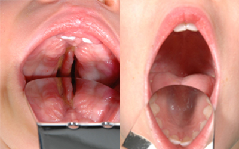
Cleft Palate Only Before and After Pictures
We treat and correct cleft lip, palate and all its variations and levels of complexity. Look at these before and after photographs to see how surgery performed by our world-class team of physicians at the International Craniofacial Institute in Dallas, Texas can improve your child’s outlook on life. Our team represents a wealth of specializations in every area related to cleft conditions: specialized craniofacial surgeons, feeding specialist, speech specialist, orthodontics and more. We work with each other, as well as with our patients families, to ensure children born with cleft/lip palate are healthy and happy. As a result, you can expect two critical outcomes – proper function and a beautifully restored appearance of all cleft-affected areas.
Cleft lip and cleft palate may be present in varying degrees, all of which require surgery. In addition, some children will have only one condition while others might be plagued with both. As such, some children may require only one surgery while others will need multiple operations to correct his or her unique differences.
Regardless of the treatment plan your child requires, he or she can obtain tremendous results from the caring, compassionate physicians here at the International Craniofacial Institute. Look at these before and after photographs of patients who have undergone cleft lip and/or cleft palate surgery to get an idea as to what results you could expect.
Cleft Palate Only
If you have a child or another family member who is suffering from a genetic syndrome or has a cleft lip, cleft palate, or craniofacial complication, the staff at the International Craniofacial Institute can help. Contact us today to talk with the doctors and staff about your options and how we can help.


