Eyes Terms
As you’ve seen happens with other sensory systems, craniofacial structural differences may affect your child’s vision throughout their growing years. If you’re familiar with the parts of that puzzle, you’ll be an assistant to us and an encouragement to your child as we take them through the needed corrections and therapy.

Amblyopia
Refers to a vision problem in which the child’s eye’s information doesn’t transmit clearly to the brain for interpretation. If this situation begins in childhood or goes on for an extended period of time, that eye becomes unable to process information at all. Most of the time, it only affects one of the eyes– often called ‘lazy eye’. Lazy eye is an inaccurate term however, since the eye muscles function normally and only image transmission is at fault.
Astigmatism
Refers to a condition in which the eye cornea or lens is misshapen, so that objects are not seen symmetrically.
Binocular Vision
Refers to the ability to use both eyes together (bini= double, oculus=eye). Binocular vision provides a 20% wider view span, helps us see faint objects, and provides more accurate depth perception than one eye alone.
Choroid
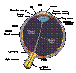
Refers to the tissue layer between the retina and sclera which is filled with blood vessels for carrying oxygen and nutrients to the outer retina.
Ciliary Muscle
Refers to the three sets of eye muscles attached to the lens, which contract or relax to shape the surface
of the lens. When relaxed, it allows the lens to flatten and see far away; when flexed, it makes the lens
more curved (convex) in order to see close objects. The muscles are three-directional to ensure equal force
on all edges of the lens: lengthwise, around the circumference, and around the entire eyeball
three-dimensionally.
Coloboma
Refers to the condition in which a person is missing a part of their eye structure due to incomplete closure of a tiny gap underneath the embryo’s developing eye. The gap (optic fissure) is allows nourishment to reach the growing eye, but must close for final eye development; if it doesn’t close, an incomplete eye results.
Concave, convex
Refers to the shape of the outside front curved surface of a rounded object. Concave refers to an
inward-facing curve; convex refers to an outward-facing curve. The eye lens may be slightly concave or
convex beyond the ideal shape, changing the way it refracts light into the back ‘seeing’ portion of the
eyeball.
Cones
Refers to special light-response, color-detection cells lining the inside central back of the eye. Called photoreceptor cells (photo=light; receptor=receives and records) — they signal the eye’s nerve cells to carry visual impressions to the brain and work especially well in bright light.
Cornea
Refers to the clear frontal eye covering which provides 80% of the eyes’ focus power; under which lies the iris (colored segment), pupil (‘camera’ opening for light), and anterior chamber (frontal inside area of the eyeball). Sensitive to touch, temperature, and chemicals, even slight stimulation of the cornea involuntarily causes the eyelid to close protectively. To assure transparency, the cornea has no blood vessels and receives oxygen directly through the air; nutrients through the eye’s outer tear fluid, inner liquid (aqueous humour), and from substances the nerve fibers transport.
Enophtalamus, Exopthalamus
Refers to either of two extremes in eyeball position within the eye sockets: deeply set eyeballs (enopthalamus); or shallowly set ‘bulging’ eyeballs (exophtalamus). In either case, orbital reconstruction can re-shape the area so as to surround the eye with the proper amount of bony protection. This repair can ease eye irritation/discomfort, provide an acceptable appearance, and improve vision.
Epibular Dermoid
Refers to a tiny growth on the eyeball surface, visible as a pinkish white raised area.
Eyelash
Refers to the tiny, even row of protective hairs lining the upper and lower rims of each eyelid. These serve to protect the eye from harmful substances: when touched even slightly by air currents, dust particles, and any kind of object, the eyelashes stimulate an immediate reflex ‘blink’, closing the eye surface off from the stimulus.
Fovea
Refers to the specific area of the center back of the retina, where your child’s most distinct and detailed vision takes place (e.g., reading, watching TV, driving, activities requiring quick hand-eye coordination. 50% of the information carried to your child’s brain about an image comes from the fovea, 50% from the rest of the retina. It appears as a tiny indentation in the eye lining (fovea=pit).
Hyaloid Artery
Refers to the part of the carotid artery which nourishes the embryo’s developing eye until about the tenth week of life after conception. At that point, the lens obtains nourishment separately, and the artery pulls back/regresses. When bits of it remain in the middle watery section of the eyeball, it can cause a patient to see ‘floaters’.

Iris
Refers to the colored light-sensitive part of the eye tissue which connects to a set of contraction and dilation muscles that act upon the eye opening (pupil). Low light stimulates the dilation muscles to allow additional light in; bright light stimulates the contraction muscles to reduce the size of the opening and allow less light into the eye. A drainage area in front of the outer iris rim maintains proper inner eye pressure; iris conditions can harm this balance and indirectly harm vision quite seriously.
Keratitis
Refers to the condition in which shallow eye socket structure or lack of adequate eyelid cover allows
frequent cornea irritation, making vision blurry and causing inflammation, pain and tearing in the
eyes themselves (kerat= cornea; it is=inflammation or irritation).
Malar surfaces
Refers to the upper heightened area of cheek bones, toward the edge of the face’s front on either side of the lower eye; the malar surface continues and forms the floor of each eye socket rim.
Myopia, Hyperopia
Refers to the two opposite conditions of an imperfectly rounded eye. In myopia (my=near, opia=sight)
the eyeball is long or the cornea is steeply shaped, focusing images in the watery inner eye chamber
instead of on the eye’s back inner wall (retina) where sight takes place. Near objects are clear,
but faraway objects are blurry and unfocused. In hyperopia (hyper=extra, long; opia=sight), the
cornea is flat or the eyeball is shortened, focusing beyond the retinal area; only faraway objects
can be properly brought into focus, and nearby objects are blurry.
Optic Disc
Refers the disc-shaped area at the back of the eye where the visual information is transferred to the optic nerve and carried to the brain. This area has no rods or cones (photoreceptor cells) and cannot respond to light; thus every eye has a ‘blind spot’ or break in their normal field of vision.
Optic Nerve
Refers to the central nerve at the back of the eye which carries visual information to your child’s brain; it is one of 12 pairs of cranial nerves but also part of the central nervous system.
Orbit
Refers to the open ‘socket areas’ in the skull’s facial area which protect and house the eyeballs. Seven different facial bones come together to form the orbit on each side of the face: the frontal, lacrimal, ethmoid, zygomatic, maxillary, palatine, and sphenoid bones.
Palpebral Fissure
Refers to the opening between the upper and lower eyelids, inside of and behind which the eyeball rests. Several syndromic craniofacial conditions can cause narrowing of this opening.
Pupil
Refers to the round center opening of the eye; with help from involuntary connected dilation and
constriction muscles, the pupil controls the amount of light allowed into the inner eye ‘seeing’
surfaces. The light entering the eye is nearly all absorbed by the inner eye tissues, so the
opening itself appears black or light-less. In normal light, the pupil is about 3-4 mm across;
in bright light it can constrict to half or less of that size; in dim light it can double its
normal size.
Retina
Refers to the eye’s inner lining of light-sensitive ‘rod’ and ‘cone’ cells, similar in function to camera film. The eye lens sends and focuses images onto the retina, stimulating nerve signals that take image information to the brain. A tiny portion of the retinal pathway responds to blue light via a substance called melanopsin, taking the information through a non-vision-related nerve to the brain to adjust the person’s awake/sleep rhythms.
Rods
Refers to the special cells that line the sides of the eye’s inner back area, respond to medium-low light levels, and translate information in shades of black and white only. After they capture the light they signal the eye nerve cells to carry the impression to the brain. These are classified as photoreceptor or light-receiving cells, as are the cones (photo=light; receptor=receives and records).
Sclera
Refers to the opaque white part of the eyeball, designed to protect the eye. It’s made of tough collagen and elastic fiber, starting out quite thin and ‘blue’ looking in infants, becoming white in childhood. It’s covered by a film called the conjunctiva.
Stereopsis
Refers to the ability of your mind to perceive a single image even though each eye sends a slightly different projection of its view to the brain (stereo= solidity, opsis=sight).
Strabismus
Refers to the condition in which a patient’s eyes do not align with one another; one or more eye
muscles don’t perform in coordination with the others (aka cross-eyes, wall-eyes). The two eyes
gaze into different places, not looking in the same direction, either due to a muscular defect
or a problem in brain-muscle information.
Strabismus means ‘squint-eyed’; a person with this condition cannot perceive depth accurately.
Suspensary Ligament
Refers to the organ that supports the eye’s lens inside of the eyeball.
Tear duct
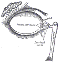
Refers to the tiny liquid-carrying channel in each eye, beginning with a minute opening at the
inside corner of each eye; extending below the skin and down the inside of the nose, creating a
fluid flow system that keeps the eyes moist, yet drained (i.e. Lacrimal duct).